MENISCUS

MENISCUS

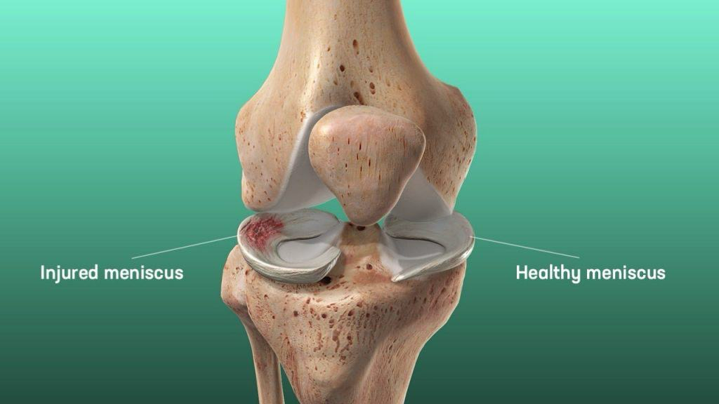
The knee joint has two menisci—the lateral meniscus and the medial meniscus. The lateral meniscus is smaller, circular, and O-shaped, while the medial meniscus is C-shaped, broader, but thinner with a flat underside. The medial meniscus is wider at the back than the front and is firmly attached to the tibia, joint capsule, and tibial collateral ligament by coronary ligaments.


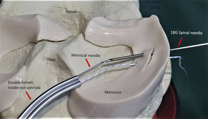
For those looking for best meniscus repair surgery , Dr Nitin Rawal and his team at AOSC specialize in advanced techniques to ensure optimal recovery and long-term knee health.

Along with meniscus repair, they also offer expertise in shoulder arthroscopy and other joint procedures for complete orthopedic care.

The lateral meniscus is loosely attached to the tibia and joint capsule, with a strong connection to the popliteus tendon at the back. Its front part merges with the ACL, and both menisci are linked by the transverse ligament in front of the ACL.

Functions:

  • Load Distribution: Helps spread weight evenly across the knee by improving joint fit.
  • Shock Absorption: More elastic than cartilage, it absorbs impact and supports 50% of weight in extension and 85% in flexion.
  • Stability: Deepens the tibial surface and acts as a secondary stabilizer, especially important after ACL injuries, where the medial meniscus plays a key role in preventing excessive forward movement of the tibia.

Meniscus Zones

  • Red Zone (Outer Third): Well-vascularized, good healing potential.
  • Red-White Zone (Middle Third): Moderate healing potential.
  • White Zone (Inner Third): Avascular, poor healing potential.

Types of Meniscal Tears

  1. Vertical/Longitudinal Tear: Most common, often seen with ACL tears.
  2. Bucket Handle Tear: A displaced vertical tear into the notch.
  3. Oblique/Flap/Parrot Beak Tear: May cause locking symptoms.
  4. Radial Tear
  5. Horizontal Tear: Common in older adults, may be linked to meniscal cysts.
  6. Complex Tear
  7. Root Tear: Functionally like total meniscectomy, always needs surgery.

Lateral Root Tear: Associated with ACL tears.

                         Medial Root Tear: Linked with chondral injuries.

Symptoms of Meniscal Tears

  • Pain on the medial or lateral joint line.
  • Locking or clicking, especially while squatting.
  • Delayed or intermittent swelling.

Physical Examination Findings

  • Joint line tenderness is the most sensitive sign.
  • Mild to moderate joint effusion may be present.

Meniscus Tear Tests

  1. Apley Compression Test:
    • Distraction Phase: The knee is flexed at 90°, and the leg is pulled up while rotating. Pain suggests ligament injury.
    • Compression Phase: Downward pressure with rotation is applied. Pain and popping suggest a meniscus tear.
    • If pain is relieved during distraction but present during compression, it indicates a meniscal tear.

Meniscus injuries require careful diagnosis and may need surgical intervention, especially in cases involving root tears or significant mechanical symptoms.

For patients seeking advanced care, Dr Nitin Rawal, regarded as the best doctor for meniscus repair, utilizes these tests as part of a comprehensive evaluation to develop precise and personalized treatment plans.

McMurray Test
• Performed with the patient lying down, the knee is bent fully and rotated.
• Medial meniscus: Knee is externally rotated while extending.
• Lateral meniscus: Knee is internally rotated while extending.
• A clicking sound or pain indicates a tear, but a negative test doesn’t rule it out.

2. Squat Test
• The patient performs deep squats with internal and external rotation.
• Pain on the inside of the knee suggests a medial meniscus tear, while pain on the outside suggests a lateral meniscus tear.

The best ACL surgeon in Delhi NCR offers state-of-the-art care for knee injuries, including treatment for a painful meniscus tear.

3. Thessaly Test
• The patient stands and rotates their knee while slightly bent at 5° and 20°.
• Pain or locking suggests a meniscus tear.

Grading of Meniscus Tears
• Grade 0: Normal meniscus.
• Grade I & II: Partial tears, not reaching the free edge.
• Grade III: Full-thickness tear involving the free edge.
Investigations
• MRI is the most sensitive test but has a high false-positive rate.
• “Double PCL” or “Double Anterior Horn” sign on MRI suggests a bucket-handle tear.

Treatment Options
Non-Surgical (First Line for Grade I & II Tears)
• Rest, NSAIDs, ice therapy, and rehabilitation.

Surgical (For Severe or Persistent Symptoms)
1. Partial Meniscectomy
• Removes damaged meniscus if repair isn’t possible (complex, degenerative, or radial tears).
• Provides short-term relief but increases the risk of arthritis over time.
2. Meniscus Repair (Preferred if Possible)
• Best for red zone (vascularized) tears, vertical/longitudinal tears, root tears, and bucket-handle tears.
• Techniques: Inside-out (strongest), outside-in, all-inside (latest and minimally invasive).

3 Meniscus Repair with ACL Reconstruction
• Healing rates are better when ACL is repaired at the same time.
• Success rate: 60% with an intact ACL, 30% if ACL is untreated.
4 Meniscal Transplantation
• Done in young patients after near-total meniscectomy (especially lateral meniscus).
• Requires 8–12 months for full recovery.
• Not recommended for arthritis, obesity, or joint instability.
5 Total Meniscectomy (Not Performed Today)
• Historically removed the entire meniscus, leading to severe arthritis.
The best approach is meniscus preservation whenever possible to prevent long-term joint damage.