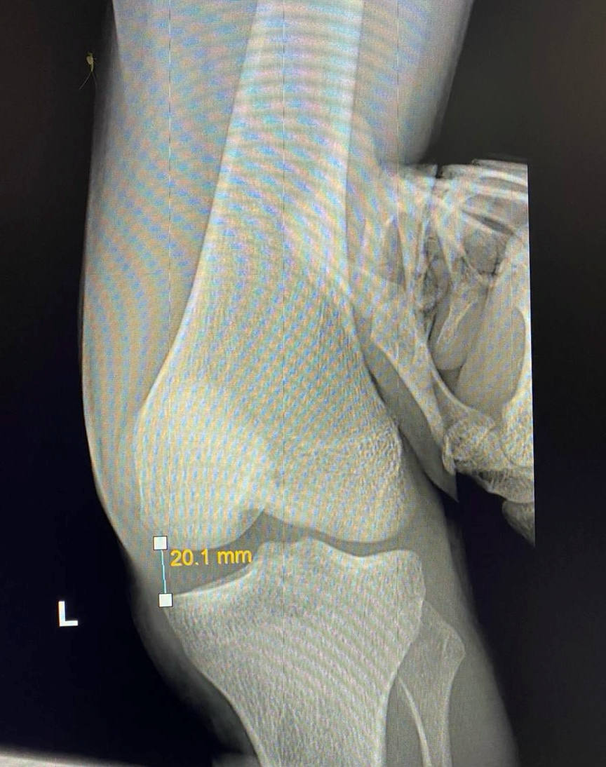
MCL

Medial Collateral Ligament (MCL) Injury

The MCL is a ligament on the inner side of the knee, connecting the thigh bone (femur) to the shin bone (tibia). It has two layers:
  • Superficial MCL – Main stabilizer against valgus stress (force pushing the knee inward).
  • Deep MCL – Supports knee stability, especially in full extension.
  • A medial collateral ligament (MCL) knee injury is a traumatic knee injury that typically occurs as a result of a sudden valgus force to the lateral aspect of the knee. Males are more commonly affected than females. Commonly occur in athletes account of 8% of all. layers of skiing, rugby, football, soccer and ice hockey have highest risk of MCL injury.

    Causes & Risk Factors
    • Direct blow to the outer knee (common in football, rugby, skiing).
    • Non-contact injuries (twisting or sudden movements).
    • Most common associated injury: ACL tear.

    Symptoms
    • Pain & tenderness on the inner knee.
    • Swelling & difficulty walking.
    • A “pop” sound at the time of injury.

    Diagnosis
    • Valgus stress test: Assesses knee laxity.

    X-ray: Usually normal but may show old injuries.
    MRI: Best for confirming MCL tears.

    Treatment

    Non-Surgical (For Grade I & II Tears Without Instability)
    • Rest, ice, painkillers, knee brace.
    Rehabilitation: Strengthening exercises for quads, hamstrings, and hips.
    Return to sports: 2 weeks for Grade I, longer for Grade II.
    Surgical Treatment (For Severe or Chronic Tears)
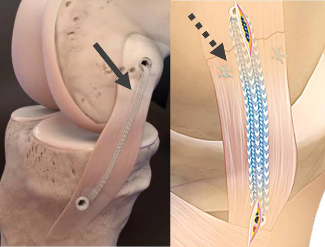
    1. MCL Repair (For fresh injuries)
    • Torn ligament is sutured back using FiberTape internal brace.
    • Faster recovery and early return to sports.
    2. MCL Reconstruction (For chronic or severe cases)
    • Hamstring graft + FiberTape for added support.
    • Minimally invasive surgery (small incisions, less pain, quick recovery).

    With modern techniques like FiberTape bracing, patients can recover faster with less pain, minimal stiffness, and early return to sports